Peripheral nerve entrapment affects millions of people, producing pain, numbness, tingling, weakness, and loss of function long after conservative care has stopped helping. Carpal tunnel syndrome is the most familiar example, but dozens of peripheral nerves throughout the body are vulnerable to compression, adhesion, and scarring. Ultrasound-guided nerve hydrodissection — the precise injection of fluid to mechanically separate a nerve from the structures pinching it — has become a meaningful option for patients caught between failed conservative treatment and surgery.
This guide is overseen by Dr. Padra Nourparvar, DO, a board-certified physician at the Stem Cell & PRP Institute of L.A. in Beverly Hills, with advanced training at CHLA, UCLA, UC Irvine, NYU, and Mount Sinai and a clinical focus on image-guided regenerative procedures. His work has been recognized with the UCLA Vice Provost Prize and the Motif Award sponsored by The Walt Disney Company, and he has cared for professional and Olympic athletes across a range of nerve and musculoskeletal conditions. The sections below explain how nerve hydrodissection works, which conditions it can address, what the published evidence shows, and where platelet-rich plasma (PRP) and stem cell–based biologics fit into the next phase of regenerative nerve care.
Amazing doctor and kind and caring staff. After my stem shot injections I had minimal pain and swelling. Highly recommend this office!
Understanding Nerve Hydrodissection
Nerve hydrodissection uses real-time ultrasound to deliver fluid precisely around an entrapped peripheral nerve, freeing it from the surrounding fascia, ligaments, scar tissue, and other compressive structures. The technique targets the root mechanics of entrapment neuropathy — pressure on the epineurium that disrupts the nerve’s own nerve supply (the nervi nervorum) and blood supply (the vasa nervorum), driving the ischemia, swelling, demyelination, and pain that patients feel.
Traditional injectates include normal saline, 5% dextrose in water (D5W), corticosteroids, local anesthetics, and hyaluronic acid. Each can provide mechanical decompression along with some anti-inflammatory or pain-relieving benefit, yet none of them actively encourages the nerve to regenerate. That gap is where regenerative biologics enter the conversation.
Why PRP and Stem Cells Change the Equation
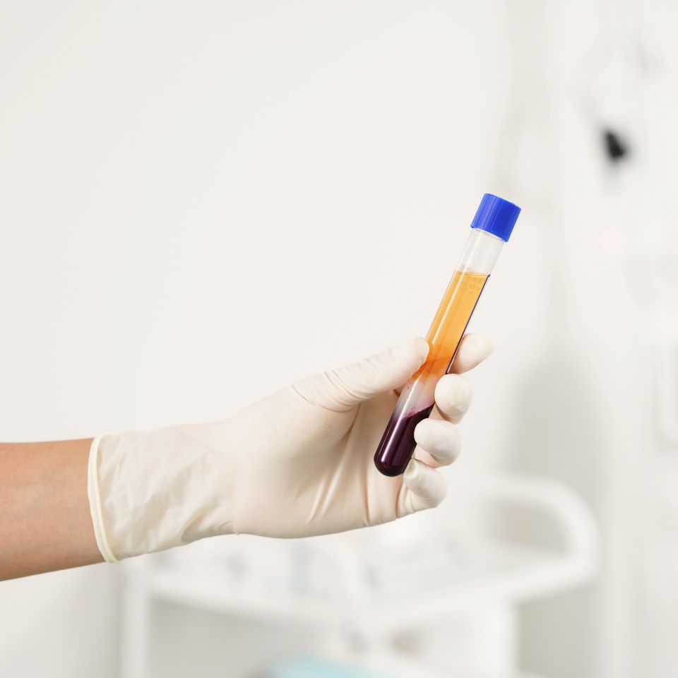
Platelet-rich plasma (PRP) is prepared from a patient’s own blood and concentrates platelets along with a cocktail of growth factors — transforming growth factor-β (TGF-β), platelet-derived growth factor (PDGF), vascular endothelial growth factor (VEGF), and insulin-like growth factor-1 (IGF-1). Used as the hydrodissection fluid, PRP works on three fronts at once: it separates the nerve mechanically, shifts healing tissue away from stiff scar toward softer and more compliant tissue, and supports nerve repair by encouraging Schwann cell activity, axonal growth, new blood vessel formation, and a calmer inflammatory response.
Stem cell therapies represent the next stage of this approach. Mesenchymal stem cells — particularly adipose-derived stem cells — and their exosomes carry neuroregenerative potential through several mechanisms: differentiation into Schwann-like cells, paracrine release of neurotrophic factors such as BDNF, GDNF, and NGF, immune modulation that shifts inflammation toward repair, angiogenesis that restores nerve blood supply, and anti-fibrotic effects that limit scarring around and within the nerve.
“Hydrodissection used to be about freeing the nerve. With regenerative biologics, the goal shifts from simply relieving pressure to giving the nerve the biological tools it needs to recover.” — Dr. Padra Nourparvar
It is worth being clear about where each therapy stands. PRP hydrodissection is performed clinically and is supported by human trials. Stem cell and exosome applications for nerve hydrodissection remain investigational and are supported largely by preclinical research — a distinction Dr. Nourparvar reviews candidly with every patient during consultation.
Conditions Treated with Nerve Hydrodissection in Los Angeles
Often overlooked in traditional practices, Platelet Poor Plasma (PPP) is the nutrient-rich fluid remaining after platelet cConditions
Treated with Nerve Hydrodissection in Los Angeles
Peripheral nerve entrapment can occur almost anywhere a nerve passes through a tight tunnel, crosses a joint, or travels near scar tissue. The conditions below span the entire body. Some are supported by randomized trials and case series, while others represent emerging or theoretical applications where the anatomy is well suited to ultrasound-guided treatment but published data is still developing.
Upper Extremity Nerve Entrapment
- Carpal Tunnel Syndrome: Median nerve compression at the wrist and the most extensively studied indication; explore carpal tunnel treatment options.
- Recurrent Carpal Tunnel Syndrome: Re-entrapment from scar tissue after a prior surgical release, where anti-fibrotic biologics are especially relevant.
- Pronator Syndrome: Median nerve entrapment at the pronator tunnel in the forearm, presenting with aching and grip weakness.
- Ligament of Struthers Entrapment: A proximal median nerve compression site above the elbow that ultrasound can reliably target.
- Cubital Tunnel Syndrome: Ulnar nerve compression at the elbow and the second most common entrapment; see cubital tunnel treatment.
- Guyon Canal Syndrome: Ulnar nerve entrapment at the wrist, anatomically analogous to the carpal tunnel; see ulnar tunnel treatment.
- Radial Tunnel Syndrome: Posterior interosseous nerve compression causing forearm pain; see radial tunnel treatment.
- Wartenberg Syndrome: A superficial sensory entrapment of the radial nerve branch in the forearm, well suited to image-guided treatment.
- Anterior Interosseous Nerve Syndrome: Forearm entrapment by fibrous bands that may respond to the anti-inflammatory and regenerative properties of PRP.
- Suprascapular Neuropathy: Compression at the suprascapular or spinoglenoid notch of the shoulder, sometimes associated with a ganglion cyst.
- Digital Nerve Entrapment: Compression of the small sensory nerves of the fingers and thumb in accessible, superficial locations.
Neurogenic Thoracic Outlet Syndrome
- C5 Nerve Root Entrapment: Prevertebral fascia release at the C5 level, studied in patients with sustained pain and disability improvement.
- Costoclavicular Compression: Entrapment near the subclavius muscle and costoclavicular space targeted across a series of hydrodissection sessions.
Brachial Plexus and Deep Structures
- Brachial Plexus Entrapment: Supraclavicular and interscalene hydrodissection used for chronic neuropathic pain of the upper limb and nerve-root region.
Spine and Nerve Root
- Cervical Radiculopathy: Cervical nerve root hydrodissection for radiating neck and arm pain; see pinched nerve treatment.
Autonomic and Sympathetic
- Stellate Ganglion Involvement: Hydrodissection performed for complex regional pain and other neuropathic pain affecting the head and neck.
Lower Extremity Nerve Entrapment
- Meralgia Paresthetica: Lateral femoral cutaneous nerve entrapment near the inguinal ligament causing outer-thigh burning and numbness.
- Posterior Femoral Cutaneous Neuropathy: A sub-gluteal entrapment described in overuse injuries and treated through perineural hydrodissection.
- Saphenous Neuropathy: Inner-knee and lower-leg pain, including infrapatellar neuralgia following knee replacement.
- Sciatic Neuropathy and Deep Gluteal Syndrome: Transgluteal sciatic hydrodissection for piriformis-related compression; see sciatica and piriformis syndrome treatment.
- Common Fibular Neuropathy: Peroneal nerve compression at the knee, including drainage of associated intraneural ganglia under ultrasound.
- Deep Fibular Nerve Entrapment: Anterior tarsal tunnel compression on the top of the foot in a superficial, accessible location.
- Tarsal Tunnel Syndrome: Posterior tibial nerve compression at the inner ankle; see tarsal tunnel treatment.
- Baxter Neuropathy: Inferior calcaneal nerve entrapment recognized as a cause of chronic, stubborn heel pain.
- Morton Neuroma: Interdigital nerve irritation between the metatarsals of the forefoot; see Morton’s neuroma treatment.
- Sural Nerve Entrapment: Outer-ankle and foot pain, including neuroma-related scarring after injury or surgery.
Trunk, Abdomen, and Pelvis
- Ilioinguinal and Iliohypogastric Entrapment: Lower-abdominal nerve pain that commonly follows hernia repair or pelvic surgery.
- Genitofemoral Entrapment: Groin and inner-thigh neuropathic pain after herniorrhaphy, appendectomy, or similar procedures.
- Intercostal Nerve Entrapment: Chest-wall neuropathic pain following thoracic surgery or rib fracture.
- Pudendal Nerve Entrapment: Deep pelvic nerve pain addressed diagnostically and therapeutically; see pudendal neuralgia treatment.
- Obturator Nerve Entrapment: Adductor-related groin pain in athletes and patients recovering from pelvic surgery.
Neuropathic Pain Conditions Beyond Entrapment
- Complex Regional Pain Syndrome: Stellate ganglion and brachial plexus hydrodissection used for persistent regional neuropathic pain.
- Postherpetic Neuralgia: Nerve pain after shingles, managed with regional and paravertebral hydrodissection approaches.
- Cervical Sprain with Arm Pain: Combined cervical root and brachial plexus hydrodissection for neuropathic upper-limb pain.
- Double Crush Syndrome: Treatment directed at the proximal compression site when a nerve is entrapped at more than one point.
- Post-Surgical Scar Entrapment: Re-entrapment from surgical scarring, where the anti-fibrotic qualities of PRP are particularly relevant.
- Intraneural Ganglion Entrapment: Ultrasound-guided aspiration of an intraneural cyst combined with hydrodissection to decompress the nerve.

The Evidence Behind PRP for Nerve Healing
Among injectable options, PRP has earned strong support in the carpal tunnel literature. In network meta-analyses of randomized trials, PRP has ranked highest for symptom relief, functional recovery, and pain reduction, ahead of D5W and corticosteroids, with SUCRA rankings reported near 91.5% for symptom relief, 92.7% for function, and 80.8% for pain. A cross-sectional cohort study found that roughly 70% of patients reported positive outcomes more than two years after a single PRP injection, with shorter symptom duration and milder nerve findings predicting better results.
Evidence extends beyond the wrist. A case series of 54 patients with radial tunnel syndrome reported pain resolution in 98% at four weeks, while a series of 14 patients with pronator syndrome showed meaningful improvement in most treated nerves at three and six months. In recurrent carpal tunnel syndrome, plasma rich in growth factors produced statistically significant improvements in pain and disability scores at 12 months. For neurogenic thoracic outlet syndrome, a study of 34 patients demonstrated sustained pain and disability improvement, and a chart review of 26 patients undergoing brachial plexus hydrodissection saw average pain scores fall from 8.3 to 1.9 at two months.
The biological rationale is consistent across these findings. PRP releases growth factors that drive tissue repair and angiogenesis, activates Schwann cells that guide regenerating axons, remodels stiff scar into more compliant tissue, modulates inflammation, and helps restore the nerve’s blood supply. Preparation matters as well — platelet concentrations of roughly four to six times baseline appear optimal, while concentrations above eight times baseline may paradoxically slow healing.
The Emerging Role of Stem Cells and Exosomes
Stem cell–based therapy is the frontier of regenerative nerve care, currently supported by a rapidly growing preclinical foundation. Adipose-derived stem cells stand out for peripheral nerve repair thanks to their strong paracrine activity, ability to take on Schwann-like behavior, low immunogenicity, and good survival after transplantation. In preclinical meta-analyses, these cells combined with nerve conduits produced the greatest improvement in functional recovery compared with other stem cell types.
Exosomes — cell-free nanovesicles released by stem cells — offer many of the same regenerative signals without the risks tied to transplanting live cells. They carry regenerative microRNAs and neurotrophic factors that encourage Schwann cell proliferation, support remyelination, and temper inflammation. In models of diabetic peripheral neuropathy, stem cell–derived exosomes improved nerve conduction velocity, increased nerve fiber density, and enhanced myelin thickness, and engineered exosomes loaded with targeted microRNAs have shown amplified effects. These applications remain experimental and are not yet approved for routine clinical use, which is why they are discussed transparently as part of a broader, individualized plan rather than as a guaranteed outcome.
What to Expect During Treatment
Nerve hydrodissection is an in-office, ultrasound-guided procedure generally considered after conservative measures such as splinting, activity modification, and medication have not delivered lasting relief, and before surgery becomes necessary. Three conditions guide candidacy: the nerve must be clearly visible on ultrasound, there must be a compressive or adhesive component to the problem, and the anatomy must allow safe needle access. Dr. Nourparvar reviews imaging, symptom history, and prior treatments to determine whether the technique — and which injectate — is appropriate.
The procedure itself is minimally invasive, and the published hydrodissection literature reports a strong safety record with no serious adverse events. Because PRP is drawn from a patient’s own blood, it carries minimal risk of allergic reaction or infection. Most patients experience only mild, short-lived soreness and resume normal activity quickly. PRP can also be a thoughtful option for patients who should avoid repeated corticosteroid injections, including those managing diabetes or blood pressure concerns.
“My job is to match the right nerve, the right anatomy, and the right biologic — and to be honest about what the evidence does and doesn’t yet show. That candor is part of good care.” — Dr. Padra Nourparvar
Frequently Asked Questions About Nerve Hydrodissection
-
It is an ultrasound-guided procedure that injects fluid around an entrapped peripheral nerve to separate it from the surrounding tissue compressing it, relieving pressure and improving the nerve’s environment.
-
A standard hydrodissection mainly frees the nerve mechanically, while PRP adds concentrated growth factors that aim to reduce scarring and support nerve repair, giving the treatment a regenerative dimension.
-
Many entrapment and neuropathic conditions may be addressed, from carpal tunnel and cubital tunnel syndrome to meralgia paresthetica, tarsal tunnel syndrome, sciatic and pudendal nerve entrapment, and more, depending on each patient’s anatomy and imaging.
-
The hydrodissection literature reports a strong safety profile, and because PRP is autologous it carries minimal risk of allergic reaction or infection; stem cell and exosome applications remain investigational and are discussed individually.
-
Most patients have only brief, mild soreness and return to everyday activity quickly, since the procedure is minimally invasive and performed without surgery or general anesthesia.
-
PRP hydrodissection is performed clinically, while stem cell and exosome-based nerve hydrodissection is experimental, supported mainly by preclinical research, and offered only within an appropriate, transparent framework.

Why Patients Choose the Stem Cell & PRP Institute of L.A.
- Ultrasound-Guided Precision: Real-time imaging allows targeted, radiation-free placement of the injectate directly around the affected nerve.
- Physician-Performed Care: Every evaluation and procedure is led by Dr. Padra Nourparvar, DO, a board-certified physician rather than a non-physician provider.
- Regenerative Focus: Treatment plans draw on PRP, stem cell, and exosome science to address the source of nerve compression, not only its symptoms.
- Comfort-Forward Technique: Needle-free Air-Jet technology and a patient-centered approach keep the experience as comfortable as possible.
- Research-Informed Protocols: Recommendations are grounded in current peer-reviewed evidence and an honest account of what each therapy can and cannot do.
- Accessible Beverly Hills Location: Care is delivered at the Cedars Sinai Medical Office Towers, serving patients from Westwood, Santa Monica, Bel Air, Brentwood, and across Los Angeles.
Stem Cell & PRP Institute of L.A.
Premier center for regenerative medicine under the leadership of Dr. Padra Nourparvar, a nationally recognized, double board-certified physician with advanced medical training from UCLA, UC Irvine, NYU, and Mount Sinai, we specialize in cutting-edge stem cell therapies designed to restore function, reduce inflammation, and elevate long-term quality of life.
Dr. Nourparvar has received numerous accolades, including the UCLA Vice Provost Prize for Best Research Article of the Year and the Motif Award for Advancement of Health from The Walt Disney Company. With over a decade of experience treating conditions such as kidney failure, stroke, Parkinson’s disease, and more, he brings a rare combination of technical skill and human compassion to every patient encounter.
Begin Your Regenerative Consultation in Beverly Hills
If nerve pain, numbness, or weakness has lingered despite conservative care, ultrasound-guided nerve hydrodissection with PRP or advanced biologics may offer a path toward genuine repair rather than another temporary fix, and a consultation is the place to learn whether it fits your anatomy and goals. To explore your options with Dr. Padra Nourparvar at the Stem Cell & PRP Institute of L.A. in Beverly Hills, book your consultation online or call or text (310) 361-5480 to schedule an evaluation.