PRP and Stem Cell Therapy for Tinnitus and Hearing Loss: What the Research Shows
Tinnitus—the persistent ringing, buzzing, or hissing in the ears—affects millions of Americans and often occurs alongside sensorineural hearing loss. These conditions can significantly impact quality of life, making everyday conversations difficult and disrupting sleep. For patients who have found limited relief from conventional treatments, regenerative medicine offers a promising new approach.
At the Stem Cell & PRP Institute of L.A. in Beverly Hills, Dr. Padra Nourparvar—a board-certified physician with advanced training from UCLA, UC Irvine, NYU, and Mount Sinai—treats patients with tinnitus and hearing loss using cutting-edge PRP and stem cell protocols. With recognition including the UCLA Vice Provost Prize for Best Research Article of the Year, Dr. Nourparvar brings both clinical expertise and research credentials to this emerging field.
Understanding How Inner Ear Damage Occurs
The inner ear contains delicate structures that are highly vulnerable to injury. Understanding these mechanisms helps explain why regenerative therapies may offer benefits that conventional treatments cannot.
Cochlear Hair Cells: These specialized sensory cells in the organ of Corti convert sound vibrations into electrical signals that the brain interprets as sound. Once damaged, cochlear hair cells do not naturally regenerate in humans—which is why hearing loss has traditionally been considered permanent.
Spiral Ganglion Neurons (SGNs): These nerve cells transmit sound signals from the hair cells to the brain. Damage to spiral ganglion neurons contributes to both hearing loss and tinnitus, as the brain attempts to compensate for reduced input.
Viral-Induced Damage: When viruses infect cochlear supporting cells, they trigger production of TRAIL (tumor necrosis factor-related apoptosis-inducing ligand), which causes hair cell death through a process called necroptosis.
Autoimmune Mechanisms: Following viral infection or medication exposure, the immune system may produce autoantibodies against inner ear structures. Autoreactive Th1/Th17 cells can attack cochlear tissue, while elevated proinflammatory cytokines (IL-1β, TNF-α, IL-6, IL-8, interferon-γ) perpetuate ongoing inflammation.
Central Neuroplastic Changes: When hair cells are damaged, the brain compensates by upregulating central neuronal gain and increasing spontaneous firing rates in auditory pathways. This hyperactivity is perceived as tinnitus—essentially the brain “turning up the volume” to compensate for reduced input.
How PRP (Platelet-Rich Plasma) Supports Inner Ear Healing
PRP is prepared from the patient’s own blood and contains concentrated growth factors that support tissue healing. Research demonstrates several mechanisms by which PRP may benefit inner ear structures:
- Promotes Spiral Ganglion Neuron Survival: PRP activates the NF-κB and p38 MAPK signaling pathways, promoting neuron survival and neurite outgrowth
- Contains Balanced Trophic Factors: These include platelet-derived growth factor (PDGF), transforming growth factor-β (TGF-β), vascular endothelial growth factor (VEGF), and insulin-like growth factor (IGF)
- Protects Outer Hair Cells: PRP demonstrates protective effects on outer hair cells in the organ of Corti and may improve auditory brainstem response (ABR) thresholds
- Promotes Cellular Migration and Regeneration: Growth factors in PRP support cellular proliferation and tissue regeneration in damaged cochlear structures
Animal Study Results for PRP Therapy
In a cisplatin-induced ototoxicity model published in The Journal of Craniofacial Surgery, intratympanic PRP demonstrated significant protective effects:
- The number of outer hair cells in the organ of Corti was significantly preserved in the PRP-treated group compared to controls
- Auditory brainstem response (ABR) thresholds showed statistically significant improvement at 3 weeks (P = 0.038)
- Morphological examination showed preservation of spiral ganglion cells and organ of Corti structure
How Mesenchymal Stem Cells (MSCs) Support Hearing Recovery
Umbilical cord-derived MSCs—from both cord tissue (Wharton’s jelly) and cord blood—work through several distinct mechanisms that address different aspects of inner ear damage.
Paracrine Effects and Immunomodulation: MSCs release growth factors and cytokines that modulate the immune system. They decrease proliferation of autoreactive Th1/Th17 cells, induce production of anti-inflammatory IL-10, and generate regulatory T cells (CD4+CD25+Foxp3+ Tregs). This immunomodulation may be particularly important for patients whose hearing loss involves autoimmune components.
Homing and Regeneration: When administered intravenously, MSCs can home to damaged cochlear tissue and migrate to the spiral ganglion area, accompanied by expression of brain-derived neurotrophic factor (BDNF).
Anti-Inflammatory Effects: MSCs upregulate genes related to immune modulation, hypoxia response, and regulation of apoptosis while downregulating genes related to synaptic remodeling. Importantly, they do not generate oxidative stress or proinflammatory cytokines (TNF-α, IL-1β, IL-6, IL-12) in the cochlea.
Transdifferentiation and Tissue Repair: MSCs can transdifferentiate into spiral ligament fibrocyte-like cells and stimulate regeneration of host cochlear cells through TGF-β signaling.
Preclinical Evidence: What Animal Studies Demonstrate
Multiple preclinical studies have demonstrated significant benefits of MSC therapy for hearing loss. The research provides important insights into potential mechanisms and expected outcomes.
Systematic Review of 12 Preclinical Studies
A meta-analysis published in Molecular Biology Reports examined 12 animal studies evaluating MSC treatment for hearing loss:
- MSC treatment resulted in a mean improvement of 15.22 dB in auditory brainstem response (ABR) thresholds
- Distortion product otoacoustic emissions (DPOAE) improved by a mean of +9.10 dB
- MSCs were derived from multiple tissue sources including bone marrow, adipose tissue, and umbilical cord blood
- Doses ranged from 4 × 10³ to 1 × 10⁶ cells
Umbilical Cord Blood MSC Study (Guinea Pigs)
Research published in Biochemical and Biophysical Research Communications demonstrated dramatic hearing restoration in deaf animals:
- Deaf animals receiving intravenous human umbilical cord blood MSCs showed significant hearing restoration
- Hearing thresholds improved from 80-90 dB (profound deafness) to 40 dB—a 40-50 dB improvement
- Histological examination showed increased numbers of spiral ganglion cells and outer hair cells
- Untreated deaf animals showed no recovery and remained at 80-90 dB thresholds
Cisplatin-Induced Hearing Loss Study (Mice)
Research published in Hearing Research found that intravenous skin-derived MSCs provided significant protection:
- MSC treatment significantly reduced hearing thresholds in cisplatin-treated mice
- Cochlear hair cells were significantly preserved
- Reduced apoptosis (cell death) in cochlear tissues as measured by TUNEL and caspase-3 staining
- Gene analysis showed MSCs downregulated genes involved in apoptosis, oxidative stress, and tissue damage
- MSCs upregulated genes affecting neural connectivity, growth factor signaling, and neurotransmitter function
Noise-Induced Hearing Loss Study (Mice)
Research published in Frontiers in Cellular Neuroscience examined MSCs derived from human umbilical cord Wharton’s jelly:
- MSCs were injected into the perilymph after severe sound trauma
- Treatment induced a moderate hearing protective effect
- MSC treatment resulted in upregulation of genes related to immune modulation, hypoxia response, mitochondrial function, and regulation of apoptosis
- Downregulation occurred in genes related to synaptic remodeling, calcium homeostasis, and extracellular matrix changes
Cord Tissue vs. Cord Blood Stem Cells: Understanding the Difference
Different stem cell sources may offer distinct advantages depending on the underlying cause of hearing loss:
- Cord Tissue MSCs: Provide strong regenerative and anti-inflammatory effects, making them particularly useful for tissue repair and cellular regeneration
- Cord Blood MSCs: May work especially well for autoimmune problems or conditions potentially related to medications or viral triggers, as they help balance the immune system
- Combination Therapy: Using both cord tissue and cord blood together may provide synergistic benefits by addressing both regenerative and immunomodulatory needs
Routes of Administration: How Treatment Is Delivered
Several delivery methods may be used, sometimes in combination to maximize therapeutic effect:
Intravenous (IV) Infusion: Stem cells are delivered through a vein, providing systemic immunomodulation and addressing autoimmune or inflammatory processes throughout the body. The IV route helps balance the whole immune system and may be particularly important when tinnitus is the primary goal. The trade-off is that cells may be utilized by inflammation elsewhere in the body before reaching the ear—which is why repeat treatments may be beneficial.
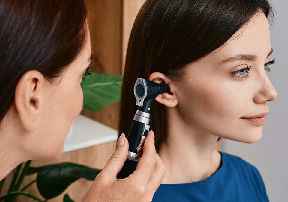
Intratympanic Injection: PRP or stem cells are injected through the eardrum into the middle ear space, where they can cross the round window membrane to reach the inner ear. This achieves higher local concentrations than IV alone.
Post-Auricular (Retro-Auricular) Injection: Injection behind the ear targets the sinus and venous drainage system that supplies the inner ear, potentially improving delivery to cochlear circulation.
Combined Approach: Using both IV and local injection provides complementary benefits—IV for systemic immune modulation and local injection for direct cochlear effects.
Why Multiple Treatments May Be Necessary
If inflammation exists in other parts of the body, the first IV treatment may be utilized addressing systemic inflammation before significant numbers of cells reach the ear. Subsequent treatments—second or third doses—can then more effectively target the ear once other inflammatory processes have been addressed. Some patients respond partially to initial treatment and more fully to subsequent sessions.
Important Considerations for Patients
Patients considering PRP or stem cell therapy for tinnitus and hearing loss should understand several key points:
- These therapies are considered investigational for hearing loss and tinnitus
- While animal studies show promising results, human clinical data specifically for tinnitus remains limited
- Results vary between individuals—some patients experience significant improvement while others may not respond
- Outcomes cannot be guaranteed
- These treatments are generally considered safe, but as with any medical procedure, potential risks exist
Dr. Nourparvar’s approach at the Stem Cell & PRP Institute of L.A. emphasizes evidence-based protocols, thorough patient evaluation, and transparent communication about expected outcomes. All procedures follow FDA-compliant guidelines and are documented in the clinic’s outcomes registry.
What to Expect During Treatment
Patients who decide to pursue treatment will receive a detailed discussion of specific dosing options, treatment schedules, and logistics during consultation. For patients traveling to Beverly Hills for treatment, planning to remain in the area for approximately one week is typically recommended. The second injection can be performed 4-7 days after the first, and patients should avoid air travel for 2-3 days after intratympanic injection.
Schedule a Consultation at the Stem Cell & PRP Institute of L.A.
For patients in Los Angeles and Beverly Hills experiencing tinnitus or sensorineural hearing loss who have not found adequate relief from conventional treatments, regenerative medicine may offer new possibilities. Dr. Padra Nourparvar provides comprehensive evaluation and individualized treatment protocols based on each patient’s specific condition and goals.
Contact the Stem Cell & PRP Institute of L.A. at (310) 361-5480 to schedule a consultation at the Beverly Hills office, located in the Cedars Sinai Medical Office Towers at 8631 West 3rd Street, Suite 545E.
References
- Chorath K, Willis M, Morton-Gonzaba N, Moreira A. Mesenchymal Stem Cells for Sensorineural Hearing Loss: A Systematic Review of Preclinical Studies. Molecular Biology Reports. 2020;47(6):4723-4736. doi:10.1007/s11033-020-05460-0.
- Choi MY, Yeo SW, Park KH. Hearing Restoration in a Deaf Animal Model With Intravenous Transplantation of Mesenchymal Stem Cells Derived From Human Umbilical Cord Blood. Biochemical and Biophysical Research Communications. 2012;427(3):629-36. doi:10.1016/j.bbrc.2012.09.111.
- Tsai SC, Lin FC, Chang KH, et al. The Intravenous Administration of Skin-Derived Mesenchymal Stem Cells Ameliorates Hearing Loss and Preserves Cochlear Hair Cells in Cisplatin-Injected Mice. Hearing Research. 2022;413:108254. doi:10.1016/j.heares.2021.108254.
- Yurtsever KN, Baklaci D, Guler I, et al. The Protective Effect of Platelet Rich Plasma Against Cisplatin-Induced Ototoxicity. The Journal of Craniofacial Surgery. 2020 Jul-Aug;31(5):e506-e509. doi:10.1097/SCS.0000000000006645.
- Warnecke A, Harre J, Shew M, et al. Successful Treatment of Noise-Induced Hearing Loss by Mesenchymal Stromal Cells: An RNAseq Analysis of Protective/Repair Pathways. Frontiers in Cellular Neuroscience. 2021;15:656930. doi:10.3389/fncel.2021.656930.
Posted on behalf of
Cedars Sinai Medical Office Towers
8631 West 3rd Street, #545E
Los Angeles, CA 90048
Phone: (310) 361-5480
Mon – Thu: 8:30am – 6:00pm
Friday: 8:30am – 4:30pm